Resources: Posters

Single-cell Multi-modal Profiling of In Vitro Neuronal Networks: Linking Morphology, Function, and Transcriptomics

July 8, 2026

International Society for Stem Cell Research

Nick Elder, PhD
Presented by:
Nick Elder, PhD
Presented at:
August 19, 2026

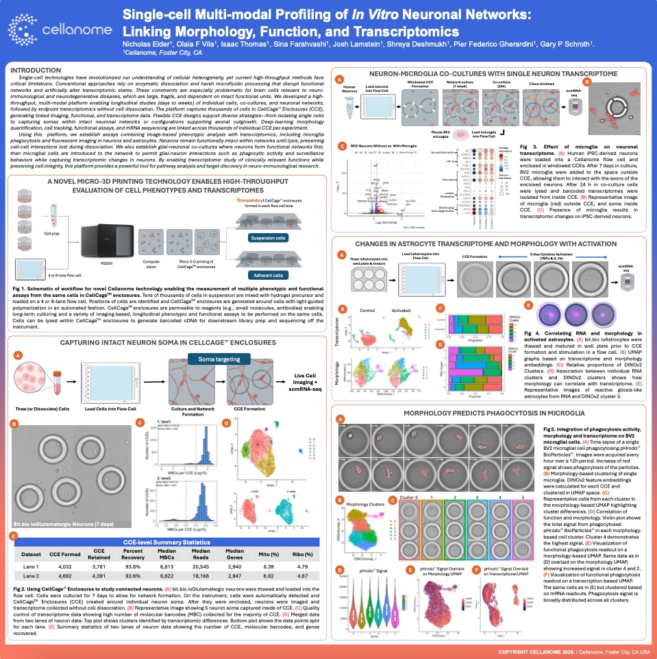
This poster presents a high-throughput workflow for studying intact neuronal networks, microglia-neuron co-cultures, and activated astrocytes using the R3200 Platform and CellCage technology. By preserving neuron soma and network connections without dissociation, the study links longitudinal imaging, functional phagocytosis assays, and single-cell transcriptomics from the same cells to reveal how morphology predicts function and how glial-neuron interactions reshape gene expression. This multimodal approach offers a powerful tool for pathway analysis and target discovery in neuro-immunological research.
Case Study: Functional Profiling of Microglia in Neuroinflammation

Link microglial behavior to gene expression at single-cell resolution, for insight into neuroinflammation, drug response, and immune dysfunction in CNS disease.

View Case Study Details

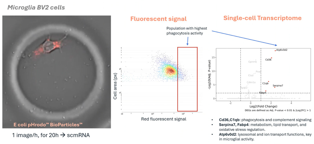
Researchers used the Cellanome R3200 to enclose individual microglia with fluorescent particles and track phagocytosis over 12 hours via fluorescent imaging. Each cell’s transcriptome was then sequenced, linking activity levels to gene expression.  

What They Found: 

High-activity microglia upregulated genes in complement signaling, lipid metabolism, and lysosomal function — key pathways in neuroinflammation and repair. 

Why it Matters: 

This approach overcomes key limitations in standard assays by capturing phagocytic function and gene expression in the same individual cells without dissociation, pooling, or inference. It enables a direct, scalable readout of immune heterogeneity, and reveals the transcriptional programs driving effective or impaired microglial responses.  

What’s Next: 

Extend to co-cultures by layering enclosed microglia over intact neuronal networks. Study how cell-cell interactions shape phagocytic behavior and fate. Combine with cytokines, CRISPR libraries, or immunotherapies to generate longitudinal datasets linking live-cell behavior with molecular readouts for MoA analysis, early biomarker discovery, and AI-guided modeling in CNS disease.

Case Study: Modeling Synapse Formation and Developmental Trajectories in 3D

Track development, function, and gene expression in intact neurospheres, a human-relevant 3D model increasingly vital as regulators move away from animal studies.

View Case Study Details

Stem-cell-derived neurospheres offer a robust 3D model of early brain development, but standard assays disrupt their structure and miss critical dynamics.  

Approach:

Using the Cellanome R3200, the research team explored:

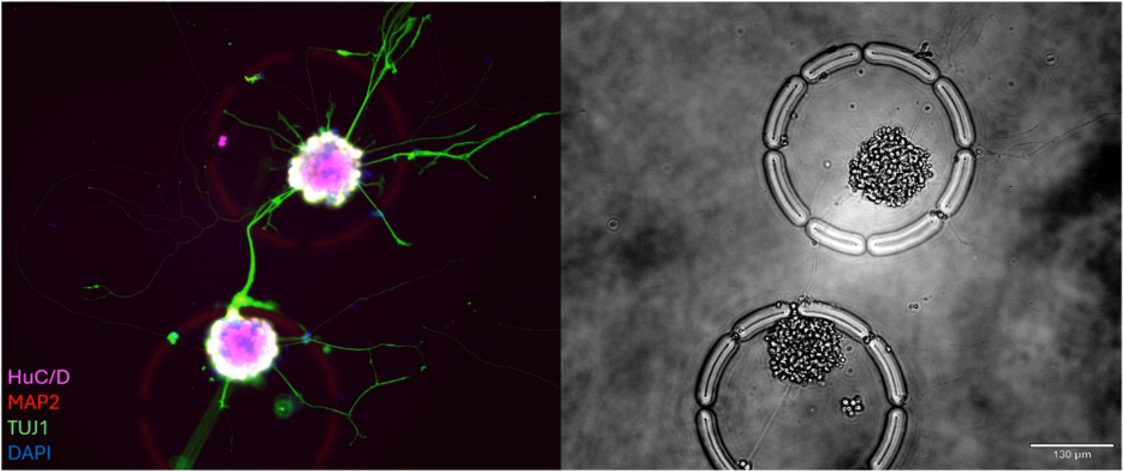
  • Hundreds of intact neurospheres (100–200 cells each) were cultured inside individual CellCage™ enclosures. 
  • Axon extension, synapse formation and calcium activity were tracked over multiple days. 
  • End-point RNA-Seq was linked back to each neurosphere’s functional behavior. 
  • UMAP clustering revealed lineage-specific gene programs, validated by fluorescent markers.  
What's Next:

This approach supports CRISPR-based screens to probe mechanisms of development, degeneration, and repair by linking perturbations to longitudinal functional and molecular readouts within preserved 3D architecture.

Why it Matters:

As the FDA and others move to reduce reliance on animal models, human-relevant in vitro systems like this are increasingly essential. 

FAQ's